
Overview: The Vital Role of Electrocardiograms in Cardiac Emergency
Discover how ECGs help diagnose cardiac emergency offering vital clues for rapid treatment and life-saving interventions in critical moments. An electrocardiogram (ECG) is a quick and simple non-invasive test that records the electrical signals of the heart. This test helps healthcare professionals diagnose a variety of heart conditions, including arrhythmias, heart attacks, and other abnormalities in heartbeat rhythms. An ECG can be performed in many settings such as medical offices, hospitals, operating rooms, and even with personal devices like smartwatches or in ambulances. For patients experiencing symptoms like chest pain, breathlessness, dizziness, fainting, or a feeling of heart racing, fluttering, or pounding chest palpitations, this test is critical. It helps doctors check if the underlying problem is related to coronary heart disease, arrhythmias, or other cardiac conditions.
ECGs are particularly useful in emergency department settings, as they provide immediate results to help diagnose cardiac emergencies. For example, ECGs can identify conditions like supraventricular or ventricular arrhythmias, heart block, and channelopathies. The electrocardiogram is crucial in detecting heart attack signs, angina, and heart failure symptoms. If the ECG records abnormal rhythms or irregular heartbeats, it can indicate the need for urgent treatment. However, while the ECG is invaluable, it has certain limitations and may not diagnose all types of heart disease, especially if normal ECG results occur. In these cases, further tests and clinical assessments are needed to confirm a diagnosis. Overall, the ECG serves as a first-step diagnostic tool that provides critical information to guide treatment in emergency situations.
What is an electrocardiogram?
Understanding an Electrocardiogram (ECG or EKG)
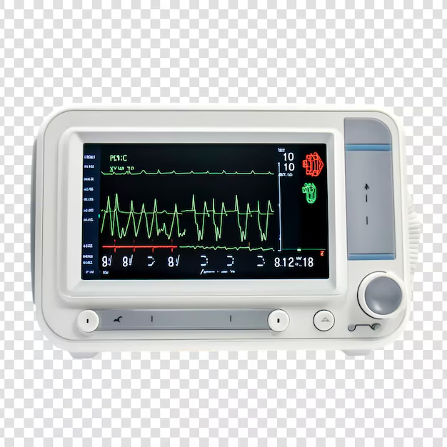
An electrocardiogram, also called an ECG or EKG, is one of the simplest and fastest tests to evaluate your heart’s electrical activity. During the procedure, small electrodes attached to plastic patches are placed on specific spots on the chest, arms, and legs. These electrodes are connected to an ECG machine using lead wires, allowing the machine to measure and record your heart’s natural electrical impulses.
These impulses, generated by your body’s own electricity, coordinate the contractions of different parts of your heart, ensuring blood flows efficiently. When the ECG records these signals, the data is interpreted, printed, and analyzed for any changes in the heart’s function. Variations in timing, whether your heart beats too fast, too slow, or in an irregular rhythm, can indicate heart-related conditions such as arrhythmias or ischemia.
My Professional Insight
In my three years as a hospital pharmacist, I’ve seen countless emergencies where a timely ECG saved lives. By detecting even slight shifts in electrical impulses, doctors can act quickly when they spot signs of heart beats being unstable or changes signaling a potential heart attack. Its ability to pinpoint irregularities, even in the different parts of the heart, ensures patients receive immediate and accurate care.
Why it’s done
The Role of Electrocardiograms in Monitoring Heart Health
An electrocardiogram (also known as ECG or EKG) is a vital tool that your care team uses to diagnose heart issues like irregular heartbeats, arrhythmias, and even a heart attack. If you’ve ever experienced chest pain, a pounding heartbeat, or symptoms like dizziness, shortness of breath, or fatigue, an ECG is often the first step in understanding the cause. This test helps detect issues such as blocked or narrowed arteries, which can reduce blood flow to the heart, or problems with a pacemaker or heart’s electrical system.
Modern ECG tools, such as a portable ECG or Holter monitor, allow your healthcare team to track your heart activity even during your daily activities. These devices can record your heart’s behavior over time, ensuring that subtle changes, like a fluttering heartbeat or skipping heartbeat, aren’t missed. Some people even use personal devices like smartwatches equipped with electrocardiogram apps to monitor their irregular heart rhythms and share data with their heart doctors. According to the American Heart Association, this individualized use of technology can provide early warnings for heart disease and improve overall treatment outcomes.
Types of ECG Tests
There are three major types of ECG tests: resting ECG, ambulatory ECG, and exercise stress test. Each serves a unique purpose in diagnosing and understanding heart conditions.
Resting ECG
A resting ECG is the most common type, where you lie down and remain still. Movement is not allowed, as it could interfere with the test by disrupting the electrical impulses the heart sends to the muscles. This test usually takes around five to 10 minutes. It’s often done to check how the heart is working properly, especially after things like a heart attack. If you’re recovering from a heart issue, the doctor may ask you to write down or record any symptoms you experience so they can be compared to the results.
Ambulatory ECG
An ambulatory ECG uses a portable recording device worn for up to 24 hours. This allows you to move around normally while your heart’s electrical activity is monitored. This type of ECG is helpful when symptoms are intermittent or stop-start, like with some arrhythmias. The doctor will arrange for you to wear this device, and you’ll be asked to write down any symptoms you experience so they can be compared to the recorded data.
Exercise Stress Test
An exercise stress test helps your doctor understand how your heart responds to physical activity. It typically involves activities like riding a stationary exercise bike or walking on a treadmill for 15 to 30 minutes. This test helps assess your heart’s response to increased physical strain, and it’s often used to determine how certain medicines or treatments might affect your heart during stress.
Syncope
Syncope: Understanding its Role in Cardiac Diagnoses
Syncope is defined as a transient loss of consciousness that occurs due to global cerebral hypoperfusion. It is often associated with postural tone loss and typically involves spontaneous recovery. This condition is a common chief complaint in both children and adolescents, and various studies have shown that syncope accounts for a significant portion of pediatric ED (emergency department) visits. In fact, syncope contributes to approximately 1% of all pediatric ED visits and has been a topic of interest in studies evaluating children and adolescents separately. While vasovagal syncope is the most frequent cause, cardiac etiologies are present in 1-6% of cases, which is why an ECG becomes crucial in evaluating these potential cardiac conditions. A careful examination through ECGs can help in diagnosing the underlying cardiac causes of syncope, distinguishing them from more benign causes, ensuring the right treatment for young patients.
Palpitations
Palpitations: A Closer Look at the Symptom
Palpitations are a subjective sensation that can occur when there is an increased frequency or strength of cardiac contractions. For pediatric patients, these sensations are commonly described in various ways, such as flipping, fluttering, beeping, or racing in the chest. It’s a common chief complaint among patients presenting to pediatric EDs (PEDs), along with other symptoms like syncope and chest pain. While palpitations are often benign, it is essential to consider the possibility of underlying cardiac issues, particularly when reported with other concerning symptoms. Palpitations in pediatric patients, although occurring in less than 0.1% of cases in some studies, may indicate more serious conditions that require closer evaluation. Importantly, an ECG is a valuable tool in diagnosing these conditions, helping determine whether the palpitations are due to an abnormal cardiac rhythm or other factors. Palpitations can sometimes be benign, but understanding the context and ensuring patients are developmentally capable of expressing their symptoms is vital for proper diagnosis and treatment.
Chest Pain
Chest Pain: When Should an ECG Be Considered?
Chest pain is a common complaint that leads many patients to seek medical attention, and it is a frequent reason for referral to pediatric cardiologists. This symptom accounts for approximately 1% of patients seen in pediatric EDs (PEDs), with cardiac etiology found in about 1-4% of these cases, according to various studies. In the ED setting, an ECG is frequently obtained as part of the initial workup for chest pain. It helps to rule out any underlying cardiac causes such as arrhythmias or coronary issues. Some tertiary care PEDs use specific algorithms to guide the ECG process, ensuring that ECGs are performed when needed and providing doctors with valuable insights. An ECG at the time of chest pain presentation can be essential for evaluating patients and ensuring they receive the appropriate care, either confirming a non-cardiac origin or identifying an underlying issue requiring urgent intervention.
Congenital and Acquired Heart Disease
When a patient is presenting with symptoms that could indicate a heart disease, an ECG becomes a critical part of the workup. An ECG helps identify abnormalities in the heart’s electrical activity, which can point to various types of congenital heart disease. These conditions are often associated with specific lesions in the heart’s structure and function. Some congenital defects might show up in the ECG as irregular heart rhythms, while others might not be as easily detected without further testing. On the other hand, acquired heart disease conditions such as pericarditis, myocarditis, or ischemia can also manifest on an ECG. These diseases, often acute or resulting from long-term conditions like rheumatic fever or rheumatic heart disease, can cause significant changes in the heart’s rhythm and electrical signals, making electrocardiography a useful tool in diagnosing these issues quickly.
Metabolic Derangements
ECG and Electrolyte Abnormalities
Metabolic derangements and electrolyte abnormalities can significantly affect the ECG. Changes in potassium, calcium, magnesium, and sodium levels are commonly observed, and the ECG can show abnormal patterns in response to these electrolyte disturbances. Clinically significant changes in the ECG are often the first sign of an electrolyte imbalance, such as hyperkalemia, which is a life-threatening condition. The first ECG change typically noted in hyperkalemia is peaking T waves, which can be recognized and treated immediately. Identifying these changes early allows healthcare professionals to address the primary electrolyte disturbance and prevent more severe complications
Toxicology
ECG and Pediatric Overdoses
In cases of pediatric overdoses, especially accidental ones, ECG monitoring plays a role in assessing potential heart-related effects. The chief complaint often includes symptoms like ingestion of a substance that may lead to cardiac abnormalities. If a child presents to the PED with signs of an overdose, contact with a poison control center is recommended for specific advice. In some cases, the ECG may have low yield in identifying issues, especially in asymptomatic patients. However, if the ingestion was of a nonaccidental nature or a specific high-risk substance, the ECG may be more useful, and it is prudent to monitor for any potential changes in the heart’s electrical activity. Age and size of the child are key factors that influence the ECG interpretation, and in such situations, it’s important to assess the overall risk and adjust care accordingly.
Is There Any Risk with an Electrocardiogram (ECG)?
Getting an electrocardiogram (ECG) is a safe and painless procedure with no risk of harm. The test doesn’t use electricity to affect your body, so there’s absolutely no chance of an electric shock. Instead, small sensors called electrodes are placed on your skin using adhesive patches. These sensors detect your heart’s natural electrical signals.
Some people might experience a slight rash or discomfort when the patches are removed, as it can feel similar to taking off a bandage. However, this sensation is brief and mild, making ECGs an easy and non-invasive way to assess heart health.
How to Prepare for an Electrocardiogram (ECG or EKG)?
You typically do not need special preparation for an electrocardiogram (ECG or EKG). However, it’s important to inform your healthcare team about all medicines you are taking, including prescription drugs, those taken without prescription, and even supplements. Certain medications and supplements may affect the test results, so your doctor may advise whether to take them as usual or pause them temporarily.
Clear communication with your healthcare team ensures the most accurate results and helps avoid potential interference during the test.
Where an Electrocardiogram (ECG or EKG) Can Be Done
An electrocardiogram (ECG or EKG) is a versatile test that can be done in various settings. It is commonly performed in a medical office or hospital, but it may be done in more urgent situations, such as in an ambulance or emergency vehicle during a cardiac emergency.
The ability to conduct ECGs in different locations ensures that patients receive timely care, whether they are in a controlled environment or experiencing a crisis on the move. This flexibility makes ECGs an essential tool in diagnosing and managing heart-related issues.
Before
Preparing for an Electrocardiogram (ECG)
Before getting an electrocardiogram (ECG), you may be asked to change into a hospital gown for easier access to the areas where the electrode patches will be applied. A member of your healthcare team might also shave any hair on the area where the patches are placed. This step helps the patches better stick to your skin and ensures accurate readings.
Once ready, you will usually be asked to lie down on an examining table or bed to allow the procedure to begin comfortably. The process is quick and non-invasive.
During
During an electrocardiogram, up to 12 sticky patches called electrodes are attached to your chest and sometimes to your arms and legs. These patches are connected by wires to a computer, which prints or displays the results as waves. These waves represent the electrical signals that travel through your heart with each heartbeat.
You will be asked to breathe normally, stay still, and do not talk during the test, as even slight movement can interfere with the accuracy of the test results. The procedure is quick, simple, and painless
After an Electrocardiogram (ECG)
Unless the test reveals a heart problem requiring immediate treatment, you can usually resume your daily activities right after an electrocardiogram. The procedure is non-invasive and has no recovery time, making it a convenient diagnostic tool.
Results
A healthcare professional will talk to you about your electrocardiogram (ECG or EKG) results, which are often available the same day as the test. These results may also be shared at your next appointment. The ECG provides critical information about heart signal patterns, including your heart rate—the number of times your heart beats per minute—and the time between each heartbeat.
An ECG is especially helpful for detecting irregularities, such as tachycardia, bradycardia, or heart rhythm disorders like arrhythmias, atrial fibrillation (AFib), and atrial flutter. It can also diagnose a current or previous heart attack, identify changes in ECG patterns caused by a damaged heart or reduced blood and oxygen supply, and detect structural changes like an enlarged heart or congenital heart defects. If a change in heartbeat or other findings is identified, your care team may recommend additional tests, such as an ultrasound or echocardiogram, to better understand your condition.
About an ECG test
How an Electrocardiogram (ECG) Helps Diagnose Heart Conditions?
An electrocardiogram (ECG) is a test used to diagnose various heart conditions by recording the electrical activity of your heart. During the procedure, small sticky dots called electrodes are placed on your chest, arms, and legs, connected by wire leads to an ECG machine or electrocardiograph. This device records the heart muscle’s electrical signals, producing a trace on a screen or paper. Any abnormality in the heart rhythm or heart rate can indicate issues like damage to the heart or poor blood supply.
The doctor will carefully examine the trace for specific features that may point to different heart conditions, such as heart attacks, abnormal heart rhythms, or arrhythmias. These include rapid, slow, or irregular heart beats, which can signal underlying problems like inflammation from pericarditis or myocarditis, or even life-threatening conditions like cardiac arrest. If you’re experiencing symptoms such as chest pain, dizziness, fainting, fatigue, heart racing, or fluttering, an ECG can help recommend the best course of action. Symptoms like shortness of breath, sweating, or palpitations, along with warning signs of a heart attack or angina, might prompt further monitoring to understand the effects and guide treatment, including medicines or implantable cardiac devices like a permanent pacemaker
Having an ECG
An ECG is a simple test that usually doesn’t require special preparation. You can eat and drink as you normally would, unless your doctor has advised otherwise. It’s important to let your doctor know if you’re taking any medications or have allergies to adhesive tapes or adhesive materials, as these might be used to attach the electrodes to your skin.
During the test, you may be asked to remove your upper clothing to allow the electrodes to be placed on your chest, arms, and legs. If you’re wearing a separate top, trousers, or skirt, it will be easier to access these areas. If you’re wearing an underwire bra, you may be asked to remove it, as it could interfere with the ECG reading. For the test to work best, your skin should be clean, dry, and free of oils or lotions. If necessary, the technician may shave any hair to ensure the electrodes stick properly
After an ECG
Once the ECG electrodes are removed from your skin, you can resume your normal activities immediately. Your doctor will usually interpret the results in the context of your medical history, symptoms, and clinical examination to determine the treatment you might need. If necessary, they will discuss the various treatments available and recommend the most suitable options for your heart condition.
A normal ECG result doesn’t always mean that your heart condition isn’t the cause of the problem. If the electrical activity of the heart appears normal but symptoms persist, your doctor may recommend other tests to investigate further. These tests may include a physical examination, listening to heart sounds, a chest X-ray to produce an image of the location, size, and shape of the lungs, heart, and major blood vessels, an echocardiogram (ultrasound), magnetic resonance imaging (MRI), CT scans for 3D images of internal organs and structures, or blood tests. In cases of suspected heart attack, a coronary angiogram can be performed to examine the coronary arteries.
Possible complications of an ECG
Safety of ECG and Its Purpose
An ECG is a very safe test that doesn’t involve sending strong electric currents through your body. The adhesive used to stick the electrodes to your skin may cause mild itchiness or redness, but this usually resolves on its own without the need for any treatment. If you experience any irritation or discomfort, you can request your healthcare provider to look for the cause.
The ECG is used to evaluate heart-related problems like chest pain, severe tiredness, fatigue, shortness of breath, dizziness, or fainting. It can identify irregular heartbeats and help assess your overall health. If necessary, the results may be used to plan procedures or surgery, such as heart surgery, cardiac catheterization, or even the implantation of a pacemaker. The ECG provides a baseline tracing of your heart’s function, which can be compared to future ECGs to monitor changes. Your doctor may advise further testing or treatment based on the ECG results, particularly if conditions like heart attack, myocardial infarction, or endocarditis (inflammation or infection of the heart valves) are suspected.
What are the risks of an electrocardiogram?
Risks and Considerations of an ECG
An ECG is a quick and easy way to assess your heart’s function, and the risks associated with the test are minimal. It is rare to experience any significant issues. Most people feel nothing or only mild discomfort from the sticky electrodes that are placed on the skin. However, if the electrode patches are left on for too long, there is a slight chance of skin irritation.
The risks can vary depending on your specific medical condition. It’s important to discuss any concerns with your healthcare provider before the test, especially if certain factors or conditions might interfere with the results. For example, obesity, anatomical considerations (such as the size or location of your chest or heart), movement (due to exercise or smoking), and even certain medicines can affect the results. Electrolyte imbalances, such as too much or too little potassium, magnesium, or calcium in your blood, can also impact the ECG and should be discussed with your provider.
How do I get ready for an electrocardiogram?
Preparation for an ECG
Before your ECG, the healthcare provider or technician will explain the test and may ask you a few questions. Generally, fasting or not eating isn’t required before the test, but you should always tell your provider about all the prescription, over-the-counter medicines, vitamins, herbs, and supplements that you take. It’s also important to mention if you have a pacemaker or any other medical devices.
Based on your medical condition, your provider may request specific preparation or modifications to the standard procedure. Always follow any special instructions given to ensure the best results.
What happens during an electrocardiogram?
The Process of Having an ECG
An ECG is typically done on an outpatient basis, but it may also be part of a hospital stay depending on your condition. The steps may vary based on your specific condition and the provider’s practices, but the general process usually follows these basic steps:
First, you will be asked to remove any jewelry or objects that could interfere with the test. You may need to change into a gown or sheet, depending on what’s comfortable and necessary to expose your skin in areas like your chest, arms, and legs. You will be asked to lie flat on a table or bed, and it’s important to remain still during the test. Talking or moving could change the results.
If you have hairy areas on your chest, the technician may shave or clip small patches of hair to ensure the electrodes can stick to your skin properly. Once the electrodes are applied, lead wires will be attached to connect the electrodes to the ECG machine’s computer, which will begin recording the heart’s electrical activity. The tracing is usually completed in a short time, after which the leads are disconnected.
What happens after an electrocardiogram?
After your ECG, you can generally resume your normal diet and activities, unless your provider tells you differently. However, if you experience any chest pain, shortness of breath, dizziness, or fainting after the test, it’s important to follow any special care instructions provided by your healthcare team. Your provider will give you instructions depending on your situation, and they will explain the next steps based on the test results.
Before agreeing to the test or any procedure, your provider should give you information about the name of the test, the reason for it, and what to expect from the results. They will also explain what the results mean, the risks, benefits, and any possible side effects or complications. If you’re unsure about anything, you can ask who will perform the test, their qualifications, and if there are any alternative procedures. If something happens that you didn’t expect or if you have any questions or problems, you should call your provider for guidance. If you’re concerned about how to pay for the test, you can ask about costs or payment options as well.