
Discover key imaging tests for cardiovascular emergencies—rapid, precise, and life-saving diagnostics for heart conditions. When a cardiovascular emergency strikes—such as a heart attack, aortic dissection, or pulmonary embolism—doctors rely on imaging tests to make rapid and accurate diagnoses. These tests help visualize the heart, blood vessels, and surrounding structures to guide life-saving treatments.
What Are Imaging Tests For Cardiovascular Emergencies?
Imaging tests are diagnostic tools that provide real-time visuals of the heart, arteries, veins, and lungs. They help detect blockages, clots, heart muscle damage, fluid buildup, and structural abnormalities. The type of test used depends on symptoms, urgency, and suspected condition.
1. Electrocardiogram (ECG/EKG) – First Step in Cardiac Emergencies
What is an ECG?
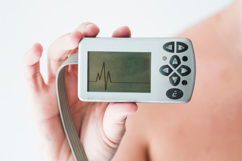
An electrocardiogram (ECG or EKG) is a non-imaging test, but it is the first-line diagnostic tool in cardiovascular emergencies. It records the electrical activity of the heart and detects life-threatening conditions such as:
- Heart attack (Myocardial infarction) – ST-elevation, T-wave inversion, Q-waves
- Arrhythmias – Atrial fibrillation, ventricular tachycardia
- Electrolyte imbalances – Hyperkalemia, hypokalemia
- Pericarditis – PR-segment depression, diffuse ST elevation
Why is an ECG Essential?
- Fast (Done in 5-10 minutes)
- Non-invasive & cost-effective
- Helps determine if more imaging is needed

2. Chest X-Ray – Quick Overview of the Heart and Lungs
What is a Chest X-Ray?
A chest X-ray (CXR) is often the first imaging test performed in cardiovascular emergencies. It uses low-dose radiation to capture images of the heart, lungs, and major vessels.
What Can a Chest X-Ray Detect?
- Heart failure – Enlarged heart, pulmonary congestion
- Pulmonary embolism – Signs of lung infarction (Hampton’s hump)
- Aortic dissection – Widened mediastinum
- Pneumothorax – Collapsed lung
- Fluid buildup – Pleural effusion, pulmonary edema
Advantages of Chest X-Ray
- Widely available & inexpensive
- Fast results (within minutes)
- Useful for ruling out lung-related causes of chest pain
3. Echocardiogram (Echo) – Ultrasound for Heart Function
What is an Echocardiogram?
An echocardiogram (echo) uses ultrasound waves to create moving images of the heart’s chambers, valves, and blood flow. It is one of the most versatile cardiac imaging tests in emergencies.
Types of Echocardiograms Used in Emergencies
- Transthoracic Echocardiogram (TTE) – Standard, non-invasive scan from the chest wall
- Transesophageal Echocardiogram (TEE) – Invasive probe inserted via the esophagus for detailed heart views
- Point-of-Care Ultrasound (POCUS) – Quick bedside echo used in the ER
What Conditions Can Echo Detect?
- Heart failure – Low ejection fraction, dilated ventricles
- Cardiac tamponade – Fluid around the heart (pericardial effusion)
- Aortic dissection – Intimal flap seen in TEE
- Valve disorders – Mitral regurgitation, aortic stenosis
Benefits of Echocardiography
- No radiation exposure
- Real-time assessment of heart function
- TEE is highly accurate for aortic dissections
4. CT Angiography – Detecting Blockages and Emergencies
What is CT Angiography?
Computed Tomography (CT) Angiography is a high-speed imaging test that uses X-rays and contrast dye to visualize blood vessels.
When is CT Angiography Used?
- Acute Coronary Syndrome (ACS) – Detects coronary artery blockages
- Pulmonary Embolism (PE) – Identifies blood clots in the lungs
- Aortic Dissection – Confirms a tear in the aorta
- Stroke Evaluation – Identifies carotid artery stenosis
Why is CT Angiography Important?
- Fast (Scan completed in seconds)
- High accuracy in detecting vascular emergencies
- Essential for surgical planning in aortic dissection
Limitations of CT Angiography
- Requires contrast dye (may cause kidney issues in some patients)
- Radiation exposure
- Not suitable for unstable patients who can’t lie still
5. Cardiac MRI – Detailed Heart Imaging Without Radiation
What is a Cardiac MRI?
A cardiac magnetic resonance imaging (MRI) scan uses powerful magnets and radio waves to create detailed images of the heart’s structure, blood flow, and tissue damage.
When is Cardiac MRI Used in Emergencies?
- Myocarditis – Inflammation of the heart muscle
- Heart attack (MI) complications – Detects scar tissue
- Aortic dissection – Alternative to CT angiography
- Cardiomyopathies – Identifies underlying structural abnormalities
Pros and Cons of Cardiac MRI
✅ No radiation exposure
✅ Highly detailed imaging
❌ Takes longer (30-60 minutes)
❌ Not ideal for unstable patients
6. Coronary Angiography – Gold Standard for Heart Attacks
What is Coronary Angiography?
Coronary angiography (cardiac catheterization) is an invasive procedure that uses contrast dye and X-rays to visualize the coronary arteries.
Why is Coronary Angiography Performed?
- Heart attack (STEMI/NSTEMI) – Identifies blocked arteries
- Angina – Determines the severity of coronary artery disease
- Prepping for angioplasty or stent placement
Key Benefits of Coronary Angiography
- Directly identifies arterial blockages
- Can be combined with stenting (PCI)
- Immediate life-saving intervention possible
Risks and Limitations
- Invasive with slight risk of complications
- Requires contrast dye (kidney concerns)
7. Nuclear Medicine Scans – Assessing Heart Perfusion
What Are Nuclear Medicine Scans?
These tests use radioactive tracers to evaluate blood flow and heart function. Common scans include:
- Myocardial Perfusion Imaging (MPI) – Assesses coronary artery disease
- Positron Emission Tomography (PET) – Detects heart muscle viability
When Are These Scans Used?
- Assessing blood flow after a heart attack
- Determining if bypass surgery is needed
What is the Right Imaging Test in Emergencies?
The choice of cardiovascular imaging test depends on the urgency, symptoms, and suspected diagnosis:
- Heart attack? → ECG + Coronary Angiography
- Pulmonary embolism? → CT Angiography
- Aortic dissection? → TEE or CT Angiography
- Heart failure? → Echocardiogram
Early detection saves lives! Understanding these imaging techniques can help doctors and patients make informed decisions during critical moments.
FAQs
1. Which imaging test is the fastest for diagnosing a heart attack?
The ECG (electrocardiogram) is the fastest test, often completed within 5-10 minutes. However, for confirming blocked arteries, a coronary angiography is the gold standard.
2. Can a chest X-ray detect a heart attack?
No, a chest X-ray cannot directly detect a heart attack, but it can help identify heart failure, fluid buildup, or lung-related issues that may cause chest pain.
3. What is the best test for detecting aortic dissection?
The most accurate tests for aortic dissection are CT angiography and transesophageal echocardiography (TEE).
4. Is an echocardiogram the same as an ECG?
No, an ECG (electrocardiogram) records the electrical activity of the heart, while an echocardiogram (echo) uses ultrasound to create moving images of the heart’s structure and function.
5. Can a CT scan detect blocked arteries?
Yes, a CT coronary angiography can detect coronary artery blockages, but a coronary angiogram (cardiac catheterization) is the most definitive test for diagnosing and treating heart blockages.
6. Is a cardiac MRI better than a CT scan?
A cardiac MRI provides detailed images without radiation, making it useful for detecting myocarditis, scarring, and structural heart issues. However, a CT angiography is faster and better suited for emergencies like heart attacks or pulmonary embolisms.
7. How long does a coronary angiography take?
A coronary angiography usually takes 30-60 minutes, depending on the complexity of the case.
8. What imaging test is best for detecting pulmonary embolism?
CT Pulmonary Angiography (CTPA) is the gold standard for diagnosing pulmonary embolism (blood clots in the lungs).
9. Do imaging tests for heart emergencies use radiation?
Yes, some tests like chest X-ray, CT angiography, and coronary angiography use radiation, while others like echocardiograms and MRI scans do not.
10. Which imaging test is safest for pregnant women in cardiovascular emergencies?
Echocardiogram (ultrasound) and MRI (without contrast) are safe options during pregnancy. CT scans and X-rays should be used cautiously.